Presumptive and Confirmatory Tests for Blood
(a) Presumptive Test
1. Phenolpthalein Test (Kastle Meyer Test)
Place a small portion of the suspected bloodstain, such as a cutting, swab, or extract, onto a piece of filter paper.
Two or three drops of ethanol are added to the stain.
Add two drops of the prepared phenolphthalein solution to the stain.
After ensuring that no color develops during this waiting period, 2-3 drops of 3% hydrogen peroxide are introduced to the stain.
The emergence of a vibrant pink color signifies a positive result for peroxide activity, indicating the presence of hemoglobin.
2. Leuomalachite Green Test (LMG)
A small curring, swab, or extract of the suspected bloodstream is placed on filter paper.
Add leucomalachite green solution to the stain.
If indicate green color appears then it gives a positive test for the presence of blood.
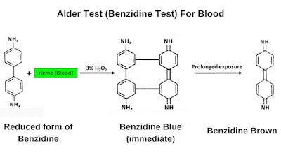
3. Alder Test (Benzidine Test)
It is the oldest method for the detection of blood which was developed by Alder in 1904.
It produces dark blue color in the reaction of blood with benzidine substrate in the presence of hydrogen peroxide.
Take a small amount of blood sample in a petri dish.
Add a small quantity of benzidine reagent to the sample, typically a few drops.
Add 3% Hydrogen peroxide to it.
Wait for 20 seconds, the appearance of dark blue color indicates a positive blood test.
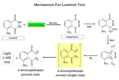
4. Luminol Test
Take the suspected blood stain and add a few amounts of Luminol reagent.
The appearance of fluorescent color indicates a positive blood test.
(b) Confirmatory Test
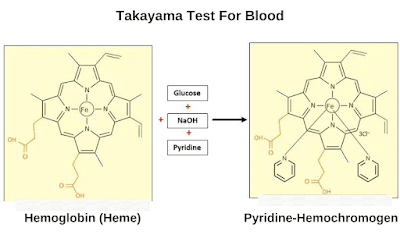
1. Takayama Test
Position the specimen to be tested onto a microscope slide and cover it using a cover slip.
Apply a single drop of Takayama reagent onto the sample, enabling it to spread beneath the cover slip.
Warm slide gently on a hot plate at 65 ℃ for 10-20 seconds
Allow to cool and observe under a microscope at 100X.
The appearance of pink needle-shaped crystals of pyridine Haemochromogen is a positive reaction for heme and conforms to the presence of Hemoglobin.
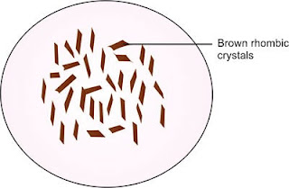
2. Teichmann Test
Position the specimen onto a microscope slide and carefully cover it with a cover slip.
Let the reagent drop under the cover slip.
Warm the slide gently on a hot plate at 65 ℃ for 10-20 seconds.
Allow to cool and observe under the microscope at 100X.
The appearance of brown rhombohedron-shaped crystals of ferriprotoporphyrin chloride is a positive reaction for heme.


